Showing posts with label Bio-Logic. Show all posts

Saturday, 20 August 2016

Would the Piano Stools please stand up?

Figure 1. A d6-piano stool complex for Iridium.

I have recently read an article from Chem. Commun. on the chemistry-biology interface [1]. It should be fascinating to both researchers in chemistry and biology alike, as the research is about the application of an artificial metalloenzyme in catalysis.

Modular in nature, the metalloenzyme consists of a host protein and the synthetic metal complex. In order to incorporate the metal catalyst to the protein host, a well-established way, for which the researchers have adopted, is the biotin-streptavidin technology.

The metal complex the researchers were using belonged to the class of d6-piano stool complex, and in this particular case, the metal was iridium (Figure 1). This type of piano-stool complexes seemed to be stable in biological environment, thus providing a benefit for the development of bio-catalysis. In order to make their catalyst more active in a biological environment, they have devised a ‘shielding strategy’, where they coated silicon nanoparticles as a protective layer on the metalloenzyme (Figure 2).

Figure 2. The design of the catalyst architecture.

The synthesis of the iridium complex involved the reaction of a protected TFA salt of the precursor di-amine with [Cp*IrCl2]2 in the presence of triethylamine at room temperature, resulting in the iridium catalyst with satisfactory yield (Figure 3).

Figure 3. Synthesis of the iridium complex.

The metalloenzyme, henceforth known as an ATHase, was covalently anchored onto the silicon nanoparticles (Figure 2). After self-assembly and the poly-condensation of silanes, a protective layer could be formed and embedded the ATHase inside the 'silica core'. The nanoparticles were visualized by scanning electron microscopy.

Figure 4. Catalysis of imine reduction by ATHase.
The ATHase was used to catalyze an imine reduction reaction (Figure 4). Depending on the specific mutant employed, it could give rise to a specific enantiomer as the product. In a nutshell, the conversion, enantioselectivity and turnover number of the silica-protected ATHase were impressive. What was notable was that, when the researchers compared the data of the catalysis of the metalloenzyme with or without silica-protection, the results were more or less comparable, signifying that the extra silica protection has not led to a negative impact to the catalysis. The silica-protected catalyst could be recycled and re-used, with almost the same enantiomeric excess in subsequent runs.

In terms of the context of the research aim, the authors would like to explore the applications of their ATHase in biological scenarios. Thus, they used the ATHase to carry out a reaction well known to anyone in biochemistry : the conversion of NAD+ to NADH. They have used absorbance spectroscopy (340 nm) – another obvious technique for biochemists – to monitor the conversion. They observed that the silica-protected ATHase had a somewhat better turnover number as compared to the non-protected version.

Another interesting aspect was that the researchers attempted to test their ATHase in vivo, which meant they did the reduction in a cellular ‘soup’. Using cell lysate of E. coli and other cellular media, satisfactory turnover numbers could be achieved, and the results were indeed better than the homogeneous metalloenzyme. They rationalized that, since the homogeneous metalloenzymes did not possess any protective layers, the catalytic activity was lost in the presence of cellular debris in the cellular ‘soup’. Thus, the silica ‘shell’ was able to protect the ATHase in the cellular media, while at the same time allowing the ATHase to carry out its mission. Even an imine reduction in urine (!!) could be achieved (TON= 4500)! 

The methodology has the added advantages that it could be conducted in physiological conditions – room temperature, pH 7, making it a stunning approach for bio-catalysis.

Impressive!

by Ed Law
20/8/2016

Reference:

1. Immobilization of an artificial imine reductase within silica nanoparticles improves its performance
Martina Hestericová, M. Rita Correro, Markus Lenz, Philippe F.-X. Corvini, Patrick Shahgaldian and Thomas R. Ward
Chem. Commun., 2016, asap
DOI: 10.1039/c6cc04604e


Wednesday, 13 July 2016

Fatten up a Few Gear

Ever since my undergraduate years in biochemistry, I have developed an intense fascination with the chemistry and biology of membranes. While most elementary textbooks on biology have often portrayed the cell membrane as a pair of lines (or curves in some cases), this organelle is actually one of the most dynamic components of the cell, and the chemistry and molecular biology behind it serve as the driving force for many wonderful molecular phenomena. 

Now, I am working in the field of bi-phasic catalysis, and it is quite gratifying to see that there are parallels between my field and that of membrane science. If possible in the future, membrane chemical biology is certainly a research field I would love to explore! This week, I have read a great article in Journal of American Chemical Society about lipid membrane and hydrogel formation [1], and I would like to share with you here.

Figure 1. Hydrogel network formation through the catalysis of a lipid membrane.


The paper is about the use of a negative-charged lipid membrane as a catalyst to facilitate the formation of a hydrogel network (Figure 1). A hydrogel is defined as a polymeric, gel-like macromolecular structure, which is made by the cross-linking of polymer chains. Hydrogels have been used in drug delivery, tissue engineering, supramolecular catalysis [2c], and many other fields.  The self-assembly and aggregation of the resulting fibrous hydrogel network has to be designed in a way so that the material properties of the resulting gel fiber can be controlled. On the other hand, the spatial position of hydrogel formation has to be carefully defined, to serve the aim of controlled release or delivery of, for example, drug molecules.

The researchers have found that a negatively-charged lipid membrane can be used as a catalyst to form supramolecular hydrogel networks. The negatively-charged liposomes can be used to catalyze the formation of a gelator molecule 3, and they have been able to achieve spatial control – the gelator molecule 3 is formed near the membrane, so that the resulting hydrogel networks can be formed in this well-defined area. The mechanism involves the generation of a local proton (H+) gradient - due to the prevalence of the negative charges. The high proton concentration facilitates the acid-catalyzed formation of hydrazonethe functional group in the gelator molecule 3.

For the chemistry, the first stage to form the hydrogel is a reaction between a hydrazide 1 and an aldehyde 2 to form the hydrazone derivative 3. At neutral pH, this reaction is very slow. Yet, the researchers have established that, when an acid or aniline is added, the reaction rate improves a lot. The structure of the hydrazone enables itself to self-assemble and forms a fiber-like structure, and aggregates towards a cross-linked network, resulting in gelation of the surrounding solvent.

The researchers are interested to see whether a negatively-charged lipid membrane can somehow catalyze the formation of hydrazone 3 and also the formation of the final hydrogel network. Their rationale is that the negative charges on the membrane can induce an increase in the proton concentration, leading to a decrease of pH and renders the chemical environment more acidic. They believe this acidic environment may catalyze the formation of the hydrazone stucture and subsequent hydrogelation.

The reaction is carried out in the following way. The hydrazide 1 and the aldehyde 2 are mixed in a defined ratio in a buffered solution at neutral pH. After that, the negative-charged liposome solution is added, and the reaction is carried out at room condition. As observed, the gelator molecule 3 is formed and eventually gelation of solvent occurs, signifying the complete formation of the hydrogel network.

How did the researchers access whether catalysis occurred from the liposome? They employed a parameter known as minimum gelation concentration (MGC). They have first measured a control value – where no liposome is added to 3 at pH 7. When a liposome from a lipid with negative charges, DPPG (Figure 2), is added to the reaction mixture with 1 and 2, a decrease in MGC is evident. Of course, if an acid or aniline is added to the reaction mixture instead, due to their catalytic effects, the MGC should also decrease. Indeed, the researchers have found that the negatively charged liposomes work even better – they lead to lower MGC as compared to the acid / aniline scenarios. As another control experiment, the addition of a positive charged liposome sample does not lead to a decrease of MGC. With the use of an UV-active hydrazide substrate, the researchers can monitor the formation of hydrazone using absorbance spectroscopy for different reaction conditions. Thus, the logical conclusion from these trials is that a negative charged membrane is essential for catalysis to occur.


Figure 2. Structure of DPPG and DOPG.

Yet, there is one important thing to note before we jump to simple conclusions. The researchers have found that, while negatively charged membrane is likely to catalyze the formation of hydrogelator network, not all negative charged membranes can achieve that. The one missing piece in the puzzle is the melting temperature, Tm, of the liposome. When the researchers added the liposome from the negative charged lipid DOPG (Figure 2, Tm -20oC, a liquid phase membrane at room temperature), to the reaction mixture, no hydrogelation occurs even at high liposome concentration. Yet, when the researchers increase the rigidity of the DOPG membrane through the addition of cholesterol (this should be familiar to anyone doing biochemistry or membrane biology), the DOPG-cholesterol hybrid membrane can then catalyze hydrogelation, suggesting the Tm of this hybrid liposome has increased.

Thus, the researchers have summarized that, in order for the liposome to catalyze hydrogelation, 2 criteria have to be fulfilled:

(1)   a negatively charged membrane surface
(2)   a solid phase at room temperature

The researchers have also established, from oscillatory rheology, that lipid concentration can control the physical properties of the hydrogel network. They also believe that liposomes are serving as nucleation points for the formation of fibrous network. I can think of a similar analogy in the case of cytoskeleton biology, where accessory proteins can serve as nucleation centers for actin polymerization.

By using confocal microscopy and a fluorescent aldehyde substrate, the researchers can also visualize the formation of the hydrogel network. When no liposomes are present, the resulting structure is very slack and un-connected. In the presence of liposomes, by contrast, the resulting hydrogel network becomes well-organized and dense, and the effect is enhanced when the liposome concentration is increased.

A very interesting aspect of the gel fiber formation occurs from the ‘underdog’, DOPG, which does not meet up to potential at the catalytic tests only until cholesterol comes to help. Rather curiously, because the Tm of DOPG is low, that meant the membrane it forms is more fluid than the membrane from DPPG. If we look at the chemical structure of DOPG and DPPG, it may shed light on this observation, and this concept is also familiar to biochemistry students. DOPG contains carbon-carbon double bonds, while DPPG is totally saturated on the carbon chains. The presence of double bonds will provide kinks and prevent a close-packing of the hydrocarbon chains, which by contrast is facile when only saturated chains are present. Thus, the unsaturated DOPG is more fluid than the saturated DPPG, and this also explains why the Tm of DOPG is lower.

DOPG has a higher affinity for the gel fibers. The affinity of the hydrogel fiber for the lipid membrane is related to the phase behavior of the hydrocarbon chains of the lipids, thus, a more fluid membrane should favor this interaction. Thus, DOPG-derived membrane seems to interact better with the hydrogel fiber than DPPG-type membrane.

What I am particularly impressed is that, by carrying out so many control experiments, the researchers draw together all the clues and provide a coherent explanation for the different performance of the DPPG and DOPG in catalysis. They propose that, because the DPPG membrane has less affinity to the hydrogel fiber, so the fiber is not blocking the way for DPPG to effect catalysis on its surface, therefore an efficient catalysis occurs and it goes on and on. In contrast, DOPG-type membrane, which is fluid and ‘loves’ the hydrogel fiber,  interacts with the gel fiber with such a high affinity that it is literally blocking the way for further rounds of catalysis. The researchers also draw analogy to the product blocking phenomena in heterogeneous catalytic systems, and I find this as an impressive explanation!

At the biological side, the researchers have also generated hydrogel fiber formations on HeLa cell systems.

All in all, this is a wonderful paper on membrane chemical biology. I have learnt a number of new techniques from it, and I am particularly impressed by the mechanistic insights, both in terms of catalytic and material, from all the great experiments they have carried out to arrive at the conclusions. Brilliant!

by Ed Law
13/7/2016

Reference:

1. Negatively Charged Lipid Membranes Catalyze Supramolecular Hydrogel Formation
Frank Versluis, Daphne M. van Elsland, Serhii Mytnyk, Dayinta L. Perrier, Fanny Trausel, Jos M. Poolman, Chandan Maity, Vincent A. A. le Sage, Sander I. van Kasteren, Jan H. van Esch and Rienk Eelkema
J. Am. Chem. Soc., 2016, asap, DOI: 10.1021/jacs.6b03853

2. Originally, I plan to talk about a self-emulsifying system which is used in the hydroformylation of lipid substrates. When I read Ref. [1], I decide to talk about that instead. It is interesting to see there are some connections regarding the two topics. Here are the references:

(a) A self-emulsifying catalytic system for the aqueous biphasic hydroformylation of triglycerides
T. Vanbésien, A. Sayede,   E. Monflier and    F. Hapiot 
Catal. Sci. Technol., 2016,6, 3064-3073
DOI: 10.1039/C5CY01758K

(b) Supramolecular Emulsifiers in Biphasic Catalysis: The Substrate
Drives Its Own Transformation
Théodore Vanbésien, Eric Monflier, and Frédéric Hapiot
ACS Catal. 2015, 5, 4288−4292
DOI: 10.1021/acscatal.5b00861

(c) Thermoresponsive Hydrogels in Catalysis
Frédéric Hapiot, Stéphane Menuel, and Eric Monflier
ACS Catal. 2013, 3, 1006−1010
dx.doi.org/10.1021/cs400118c 


Sunday, 28 June 2015

Hey Geisha, Hold on !

Figure 1. Taken from Ref. [1].

The Angew. Chem. paper I am sharing with you this time is impressive, it is on the chemistry-biology interface. [1] It is about bio-conjugation , which is applying a chemical reaction to a biological macromolecule. Let’s deal with both fields in detail, one by one.

Bioconjugation is useful, because we can start to attach unnatural or unconventional chemical substrates onto biological molecules, such as proteins, complex carbohydrate architectures or nucleic acid-based compounds. While these methodologies have found great uses in synthetic biology, the design of the reactions is often challenging. Unlike conventional organic substrates, the reactants we are dealing with here are biological molecules, which are sensitive to high temperature, extreme pH, and possess loads of stereogenic centers. Conventional wisdom shows us that we can lower the activation energy barrier of a reaction (i.e. increasing the reaction rate) by increasing the reaction temperature. However, this does not help in the case of biological molecules, as the high temperature, or a sudden perturbation in pH, can lead to negative results, such as the denaturation of proteins or possible racemizations. So, the best methods for bioconjugation should work optimally at physiological temperature (37oC) , narrow pH range, and also work well in an aqueous media.

How can we incorporate unnatural chemical functionalities into a supposedly natural biomolecule? Molecular biology methods, of course! In this case, a protein was expressed with an unnatural amino acid (UAA), with a carbon-carbon triple bond (alkyne) on it. The alkyne can act as a ‘handle’ for other reactions, and in the past, click chemistry and Sonogashira coupling has already been successfully employed.

Figure 2. The Glaser-Hay coupling reaction.

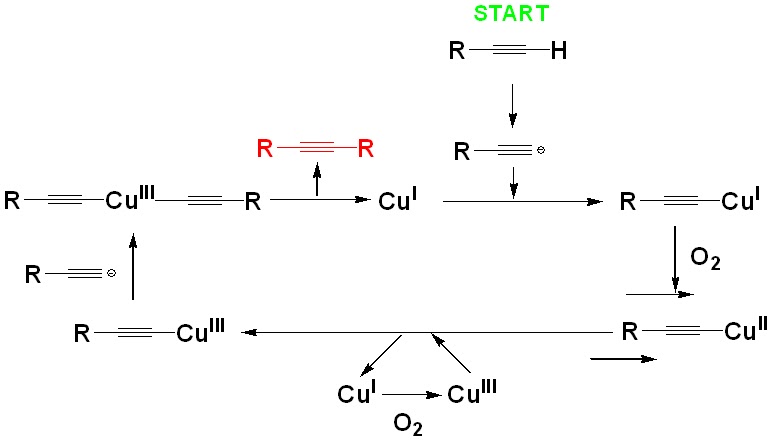
Figure 3. The catalytic cycle of Glaser-Hay coupling reaction.

The reaction in focus is known as the Glaser-Hay coupling reaction (Figure 2). It is a copper-catalyzed coupling reaction between 2 carbon-carbon triple bonds. Previous studies have concluded that the reaction probably involved a dimeric copper acetylide intermediate, and the mechanism involved an oxidation of the copper species, from +1  to +2 and then +3, and finally back to +1, continuing the catalytic cycle (Figure 3). Traditionally, this was a homo-coupling reaction, which meant you had 2 identical coupling partners to get the dimeric, symmetrical product. This unfortunately hinders its potential applications. Because what we want is a CROSS-coupling (hetero-coupling) reaction, in which you have 2 different coupling partners, A and B, to form an A-B type product (rather than an A-A or B-B type product). How about adding 2 different substrates to the Glaser-Hay system? Nope, it does not work well, as we almost alyways have a statistical amount of homo-coupling products, and that hinders purifications too. There is good news, though, as recently novel systems have been developed to lead predominately to hetero-coupled partners. For example, Lei and Agrofoglio independently developed co-catalyzed system involving a Ni (II) / Cu (I) system, and these methods work well for a variety of alkynic substrates. On the other hand, if one of the coupling partners is attached to a solid support, heterocouplings also predominate. Glaser-Hay reactions work well in organic solvents, yet it is rather unprecedented to work in an aqueous media. This article shows us that possibility.

Figure 4. The substrate for the coupling reaction. Taken from Ref. [1].

The researchers started with some model reactions on small molecules before embarking on biomolecules (Figure 4, a). With phenylacetylene and propargyl alcohol respectively, they could achieve respectively homo-coupled products in an aqueous solution with CuI / TMEDA as the catalytic system. They had to premix these 2 reagents to establish the copper complex before it could be bound to an alkyne substrate and start catalysis (Figure 5).

 Figure 5. The first stage of the catalytic cycle. Taken from Ref. [1].

To achieve unconventional biological products for their methodolgy, the researchers installed an unconventional amino acid, ‘propargyl phenylalanine’, at position 151 of the green fluorescent protein substrates they used, which was known as pPrF-GFP (Figure 4, b). They coupled this substrate with ‘AlexaFluor 488 modified alkyne’, and succeeded in making the hetero-coupled product (Figure 6, 7). They have also explored the substrate scope of their method. For solid-supported reactions, they have achieved similar yields with a propargyl or a hexyl-derived alkyne, and also in other types of biologically-related substrates (Figure 8). The results were easily visualized, as a successful coupling reaction would lead to an observable fluorescent signal, or by SDS-PAGE analysis (Figure 7).  

Figure 6. Glaser-Hay bioconjugation. Taken from Ref. [1].


Figure 7. The results of the Glaser-Hay bioconjugations as illustrated by a normal SDS-PAGE gel and a fluorescent version. In diagram A, we can see that the unconventional amino acid, pPrF, has been successfully incorporated into the GFP (Lane 2), because there is no GFP spot in Lane 3, when pPrF is absent. In diagram B, we can observe that the coupling reaction has been successful carried out because in Lane 2 and 4, which corresponds to 2 different temperatures, and with both copper catalyst and pPrF-GFP present, this substrate is coupled to the fluorophore ‘Alexa’. Therefore, fluorescene signals are evident in those lanes, signifying successful C-C bond formation. This is not the case for the other control experiments (Lane 3, 5, 6, 7), where one or both of the aforementioned components are absent. When the researches use a wild-type GFP – that means one that has no pPrF incorporated, of course no coupling occurs.


Figure 8. Other substrates. Taken from Ref. [1].

The researchers have discovered a possible unproductive side reaction - a Cu (I) -catalyzed oxidative degradation. Another interesting observation was that the reaction rates were comparable at a low (4 oC) and high temperature (37 oC). They attributed this to a  protein degradation at the higher temperature, inhibiting reaction progress.


Some personal opinions

This paper is absolutely smashing, and I hope there will be further developments on the project. In the paper, the researchers have employed 2 different alkynic substrates, a propargyl and a hexyl one. I wonder if there will be any different, in terms of reaction rates, as the chemical environment next to the 2 respective triple bonds are surely distinctive – in the propargyl case, the triple bond and the oxygen is only separated by a CH2 group, while in the hexyl alcohol case, the oxygen and the triple bond were separated by a much longer spacer. A further question would be, can we use a more hindered propargyl-type secondary ether, as we may even generate a possible stereogenic center close to the biological site (Figure 9)? The ‘double conjugated alkyne’ should serve as a nice handle for further reactions to occur – how about a thiol-yne coupling or a nucleophilic addition of ROH onto the terminal alkyne?

Figure 9. A potential asymmetric Glaser-Hay coupling reaction?

The second question - is it possible to use a Cu (II) salt, such as CuSO4 , instead to effect the coupling reaction? Obviously, this would not have the same mechanistic similarity like the Glaser-Hay type reaction, yet homocoupling involving Cu (II) salts do exist, e.g. Cu(OAc)2 for Eglinton reaction. If this is OK, then it will increase the versatility of the reaction as the Cu (II) salts are readily accessible.

It would be really exciting if this research topic can be developed much further!

by Ed Law
28/6/2015


Reference:

1. Development and Optimization of Glaser–Hay Bioconjugations
J. S. Lampkowski, J. K. Villa, T. S. Young, and D. D. Young
Angew. Chem. Int. Ed., 2015, asap.
DOI: 10.1002/anie.201502676


Tuesday, 28 April 2015

Smooth the Patchy Hedgehog


Do you know what is common between the 2 pictures shown above? They are both called ‘Sonic Hedgehog’! That’s true – in biochemistry, there is a signaling pathway known as the ‘Hedgehog Pathway’, and the proteins involved have funny names like ‘Sonic hedgehog’, ‘Smoothened’ and ‘Patched’. The April issue of Nature Chemical Biology has a really great perspective article on this pathway and the drug development involving this cell signaling circuit. [1] Indeed, Smoothened is an oncoprotein , which is coded by an oncogene. Oncogenes are obviously important because they are the ones which are overexpressed in pathological situations and ultimately lead to many type of cancer. The Hedgehog pathway is an very important signaling pathway, as it is actively involved in development. Of course, it is not at all an issue when you are developing, e.g. a fetus, but it is a big problem when you are mature. If these genes are overexpressed, it is a sort of ‘over-reaction’ and the ultimate result is malignancy. It has been shown that the abnormal signaling of Hedgehog pathway can lead to basal cell carcinoma and also some childhood tumors. This article has a bit of molecular biology and chemistry, and should be really useful for biochemistry and molecular biology students.

A diagram explaining the Hedgehog Signalling Pathway. Taken from [2].

I would also like to provide a brief discussion about the Hedgehog pathway here.  At the start, the protein ‘Patched’ inhibit the activity of ‘Smoothened’, which is a 7-transmembrane spanning, GPCR like protein. After the Hedgehog protein is cleaved and covalently modified by the addition of a palmitoyl and a cholesterol group ( both are fatty groups), that becomes hydrophobic and so it can associate with the membrane and its diffusion ability  will be limited as a result. This is indeed important because it has be previously shown that a reasonable amount of Hedgehog protein has to be present to effect downstream signaling, and so the limited diffusion  will help to establish a concentration of the Hedgehog protein.  Now, before we understand what the Hedgehog protein is up to, let us divert ourselves for one further detail. In the cell there exists a Hedgehog signaling complex (HSC), which is a series of protein that is associated with the microtubule in the cell. Among those proteins is a transcription factor called Ci, and when Ci is cleaved, it moves into the nucleus and lead to a reduced transcription of relevant genes.  However, when Hedgehog protein is present, it binds to Patched, which no longer inhibits the Smoothened Protein. To state it another way, the Hedgehog protein ‘releases’ the Smoothened protein indirectly. Smoothened is then phosphorylated by Protein Kinase A and Casein Kinase 1. Phosphorylations also occur for the other proteins in HSC and they associate with the  phosphorylated Smoothened.  Eventually, the HSC complex is no longer associated with the microtubule, and Ci is not cleaved. Therefore,  the intact Ci cannot repress transcription as a result. That is why Smoothened is an oncoprotein – because it leads to an intact Ci, which is not able to put a brake on the transcription. The wheel goes on and then ‘abnormal’ proteins are over-produced as a result. Given that ‘Smoothened’ and ‘Patched’ are nemesis, it won’t be too wrong to say ‘Smooth the Patchy Hedgehog’!

If you are interested, you can explore more about this in any biochemistry textbooks - a reasonably great understanding has been developed about this fascinating pathway.

by Ed Law
28/4/2015

Reference:

1. Regulation of the oncoprotein Smoothened by small molecules
Hayley J Sharpe, Weiru Wang, Rami N Hannoush & Frederic J de Sauvage
Nature Chemical Biology, 2015, 11, 246-255. 
doi:10.1038/nchembio.1776

2. http://www.novusbio.com/hedgehogpathway.html


Wednesday, 11 February 2015

Some Invaders (9/2/2015)

(Picture taken from http://www.cnb.csic.es/~meetinginvadosome/scientific-progamme.html.)


The concept of invadosomes in Molecular Cell Biology.


I am quite excited to hear about the concept of 'invadosome' in cell biology today. It is a set of spectacular cellular apparatus which acts as cell contacts when either some types of normal cells or tumor cells want to invade other cells, that is why it is important. The key component of the invadosome is related to its cytoskeletal machineries (in this case the F-Actin). When cellular signals, normal or nefarious, stimulate the corresponding cells, the assembly of F-actin takes place and the action is enhanced by a whole series of cytoskeletal proteins. Since focal adhesion is highly important for holding the cells together, it is not surprising at all to learn that focal adhesion kinase (FAK) will enhance the cytoskeletal action here. The completion of F-actin assembly will mobilize a whole series of extracellular matrix (ECM) - degradation machineries (such as metalloproteinases) - which will then chew up the web-like structure of the extracellular matrix. 

A personal observation from this insightful poster is that - the invadopodia (i.e. the bad guy which is related to cancer) is more 'sinister' than you bet. From the morphological diagrams, you can see that the structure of the invadopodia is far more angular and tight-knitted than the 'good guy' known as podosome. This appears to me that the invadopodia is 'ready to shoot', as an angular stance will provide a better focal adhesion strategy. This is indeed supported by the observations that these invadopodias are larger in size, they have longer persistence and have superior caliber in degrading ECM. Another fascinating question is - is there a dynamic equilibrium between the 2 types, i.e. can they interconvert? If the answer is yes, we will have to find out the conditions as this will be an important issue! A fascinating concept!

by Ed Law
9/2/2015

For the poster, please see:

Reference:

1. http://jcs.biologists.org/content/122/17/3009/F1.poster.jpg
2. http://www.cnb.csic.es/~meetinginvadosome/scientific-progamme.html